
준비 및 보관
권장 분석 절차
BD™ CompBeads can be used as surrogates to assess fluorescence spillover (Compensation). When fluorochrome conjugated antibodies are bound to BD CompBeads, they have spectral properties very similar to cells. However, for some fluorochromes there can be small differences in spectral emissions compared to cells, resulting in spillover values that differ when compared to biological controls. It is strongly recommended that when using a reagent for the first time, users compare the spillover on cells and BD CompBead to ensure that BD CompBeads are appropriate for your specific cellular application.
제품 고시
- This reagent has been pre-diluted for use at the recommended Volume per Test. We typically use 1 × 10^6 cells in a 100-µl experimental sample (a test).
- Caution: Sodium azide yields highly toxic hydrazoic acid under acidic conditions. Dilute azide compounds in running water before discarding to avoid accumulation of potentially explosive deposits in plumbing.
- An isotype control should be used at the same concentration as the antibody of interest.
- Alexa Fluor® is a registered trademark of Life Technologies Corporation.
- Please refer to http://regdocs.bd.com to access safety data sheets (SDS).
- Please refer to www.bdbiosciences.com/us/s/resources for technical protocols.
- This product is provided under an Agreement between BIOTIUM and BD Biosciences. This product, and only in the amount purchased by buyer, may be used solely for buyer’s own internal research, in a manner consistent with the accompanying product literature. No other right to use, sell or otherwise transfer (a) this product, or (b) its components is hereby granted expressly, by implication or by estoppel. This product is for research use only. Diagnostic uses require a separate license from Biotium, Inc. For information on purchasing a license to this product including for purposes other than research, contact Biotium, Inc., 3159 Corporate Place, Hayward, CA 94545, Tel: (510) 265-1027. Fax: (510) 265-1352. Email: btinfo@biotium.com.
관련 제품

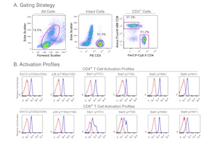
Stat (Signal transducer and activators of transcription) proteins mediate the biological activity of cytokines, including interleukins, interferons, erythropoietin, and growth factors. Ligand-receptor interaction activates constitutively-associated JAK family kinases and subsequent recruitment/activation of Stat proteins by tyrosine phosphorylation. Active Stat proteins then move to the nucleus to promote transcription of cytokine-inducible genes. Seven Stat proteins have been cloned, each of which is differentially expressed and/or activated in a cytokine-specific and cell type-specific manner. Stat5 has been characterized and shown to be encoded by two separate genes, Stat5a and Stat5b, which share over 90% identity at the amino acid level. Stat5a has been shown to be involved in lactogenesis and mammary development, while Stat5b has been shown to be involved in growth hormone signaling and liver gene expression. Both Stat5a and Stat5b are involved in IL-2 induced peripheral T cell proliferation. The peptide hormone, prolactin, binds to the prolactin receptor (PRLR) to initiate the lactogenic response. There are at least three forms of PRLR; however, only the long form activates the 92-kDa Stat5 protein by inducing phosphorylation at Y694. Once phosphorylated, Stat5 becomes an essential transcription factor which binds to the β-casein gene promoter. The presence of an SH2 domain within Stat5 suggests that it may directly interact with protein tyrosine kinases (PTKs) such as JAK2.
The 47 monoclonal antibody recognizes the phosphorylated Y694 of Stat5a. The homologous phosphorylation site in Stat5b is Y699.
The antibody was conjugated to BD Horizon Red 718, which has been developed exclusively for BD Biosciences as a better alternative to Alexa Fluor® 700. BD Horizon Red 718 can be excited by the red laser (628 – 640 nm) and, with an Em Max around 718 nm, it can be detected using a 730/45 nm filter. Due to similar excitation and emission properties, we do not recommend using R718 in combination with APC-R700 or Alexa Fluor® 700.

개발 참고 자료 (8)
-
Bromberg J, Darnell JE. The role of STATs in transcriptional control and their impact on cellular function. Oncogene. 2000; 19(21):2468-2473. (Biology). 참조 보기
-
Gouilleux F, Wakao H, Mundt M, Groner B. Prolactin induces phosphorylation of Tyr694 of Stat5 (MGF), a prerequisite for DNA binding and induction of transcription. EMBO J. 1994; 13(18):4361-4369. (Biology). 참조 보기
-
Johnston RJ, Choi YS, Diamond JA, Yang JA, Crotty S. STAT5 is a potent negative regulator of TFH cell differentiation. J Exp Med. 2012; 209(2):243-250. (Clone-specific: Flow cytometry). 참조 보기
-
Liu KD, Gaffen SL, Goldsmith MA. JAK/STAT signaling by cytokine receptors. Curr Opin Immunol. 1998; 10(3):271-278. (Biology). 참조 보기
-
Prlic M, Bevan MJ. Exploring regulatory mechanisms of CD8+ T cell contraction. Proc Natl Acad Sci U S A. 2008; 105(43):16689-16694. (Clone-specific: Flow cytometry). 참조 보기
-
Suni MA, Maino VC. Flow cytometric analysis of cell signaling proteins. Methods Mol Biol. 2011; 717:155-169. (Clone-specific: Flow cytometry). 참조 보기
-
Van De Wiele CJ, Marino JH, Murray BW, Vo SS, Whetsell ME, Teague TK. Thymocytes between the -Selection and Positive Selection Checkpoints Are Nonresponsive to IL-7 as Assessed by STAT-5 Phosphorylation. J Immunol. 2004; 172(7):4235-4244. (Biology). 참조 보기
-
Wakao H, Gouilleux F, Groner B. Mammary gland factor (MGF) is a novel member of the cytokine regulated transcription factor gene family and confers the prolactin response. EMBO J. 1994; 13(9):2182-2191. (Biology). 참조 보기
Please refer to Support Documents for Quality Certificates
Global - Refer to manufacturer's instructions for use and related User Manuals and Technical data sheets before using this products as described
Comparisons, where applicable, are made against older BD Technology, manual methods or are general performance claims. Comparisons are not made against non-BD technologies, unless otherwise noted.
For Research Use Only. Not for use in diagnostic or therapeutic procedures.
23-22944-00