-
Reagents
- Flow Cytometry Reagents
-
Western Blotting and Molecular Reagents
-
Flow Cytometry Reagents
- Immunoassay Reagents
- Single Cell Multiomics Reagents
-
Cell Preparation
-
Functional Assays
-
Microscopy and Imaging Reagents
- Western Blotting And Molecular Reagents
- Cell Preparation Separation Reagents
- Functional Cell Based Reagents
- Microscopy Imaging Reagents
- Single Cell Multiomics Reagents
- Single Cell Multinomics Reagents
-
Protocols
- BSB Protocol
-
Setting Compensation Multicolor Flow
-
Tissues Section Stain
-
Immunomicroscopy
-
Immunohistochemical
-
Immunofluorescence
-
Frozen Tissue
-
Parafin Sections
-
Fix Perm Kits
-
Protocol Direct Immunofluorscence Staining
-
Uses of Fc Block
-
Stain Lyse Wash
-
Stain Lyse No Wash
-
Mouse Splenocytes
-
Mouse Rat Leukocytes
-
Isotype Control
-
Indirect Staining Mononuclear Cells
-
Immunopurification
-
Human PBMCs
-
Human Whole Blood Samples
-
Escapee Phenomenon
-
Agarose Conjugates
-
Anti Phosphotyrosine Biotin Conjugates
-
Soluble Antibodies
-
Rabbit Polyclonal Antibodies
-
Monocloncal Antibodies
-
Horseradish Peroxidase
-
Certified Reagents
-
Biotinylated Antibodies
-
Agarose Conjugates X712261
-
Surface Staining
-
Platelet Activation
-
Intracellular Staining
-
Indirect Immunofluorescence
-
Mouse Ige
-
Cytokine Elisa
-
Induction Fas
-
Induction Dx2
-
Apoptosis By Treatment Staurosporine
-
Cell Death
-
Apo Brdu
-
Apo Direct
-
Human Cyclins
-
Detection Ki 67
-
Brdu Detection
-
Targeted mRNA Protocols
-
WTA Protocols
-
360040667732 Protocols
-
360023293831 AbSeq Protocols
-
360039007471 VDJ CDR3 Protocols
-
Annexin V Staining Protocol
-
Western Blotting with Horseradish Peroxidase Conjugates or Alkaline Phosphatase Conjugates
-
Tissue Preparation for Surface Antigen Staining
-
Account Support
-
Account FAQs
- Account FAQ Answer 1
- Account FAQ Answer 2
- Account FAQ Answer 3
- Account FAQ Answer 4
- Account FAQ Answer 5
- Account FAQ Answer 6
- Account FAQ Answer 7
- Account FAQ Answer 8
- Account FAQ Answer 9
- Account FAQ Answer 10
- Account FAQ Answer 11
- Account FAQ Answer 12
- Account FAQ Answer 13
- Account FAQ Answer 14
- Account FAQ Answer 15
- Account FAQ Answer 16
- Account FAQ Answer 21
- Create Account
- Manage Account Settings
-
PrivacyPolicy
-
Terms and Conditions
-
Account FAQs
-
- Account FAQ Answer 1
- Account FAQ Answer 2
- Account FAQ Answer 3
- Account FAQ Answer 4
- Account FAQ Answer 5
- Account FAQ Answer 6
- Account FAQ Answer 7
- Account FAQ Answer 8
- Account FAQ Answer 9
- Account FAQ Answer 10
- Account FAQ Answer 11
- Account FAQ Answer 12
- Account FAQ Answer 13
- Account FAQ Answer 14
- Account FAQ Answer 15
- Account FAQ Answer 16
- Account FAQ Answer 21
- Korea (English)
- Korea (Korea)
-
국가 / 언어 변경
Old Browser

Immune Deficiencies
The immune system provides surveillance and offers protection from pathogenic invaders and prevents detrimental responses against the self that could lead to autoimmunity. Alterations in the innate or adaptive arms of the immune system can lead to immune deficiencies. Persistent lack of adequate immune responses can open the way to chronic inflammatory conditions, which can add another layer of complexity to an already overwhelmed immune system.
Immune disorders versus immune deficiencies
Immune disorders are alterations or dysregulations of components of the immune system, be it in the immune cells or their signaling pathways. These alterations can cause either low- (immune deficiency) or hyper-activity (autoimmunity) of the immune system.
Immune system deficiencies occur when immune responses fail to protect the host against infections.
Primary immune deficiencies (PID), such as severe combined immunodeficiency (SCID), occur when some parts of the immune system are absent or deficient. PIDs are usually congenital, deriving from hereditary genetic defects.1
Secondary immune deficiencies (SID) are induced by extrinsic factors in an immune system that is intrinsically competent. Common causes of secondary immune deficiencies include immunomodulatory drugs (e.g., glucocorticoids), malnutrition (e.g., vitamin D deficiency) and chronic infection. Secondary immune deficiencies are more common than primary immune deficiencies; the most prominent secondary immune infection is acquired immune deficiency syndrome (AIDS), which results from infection with human immunodeficiency virus (HIV).


Biology of immune deficiency diseases
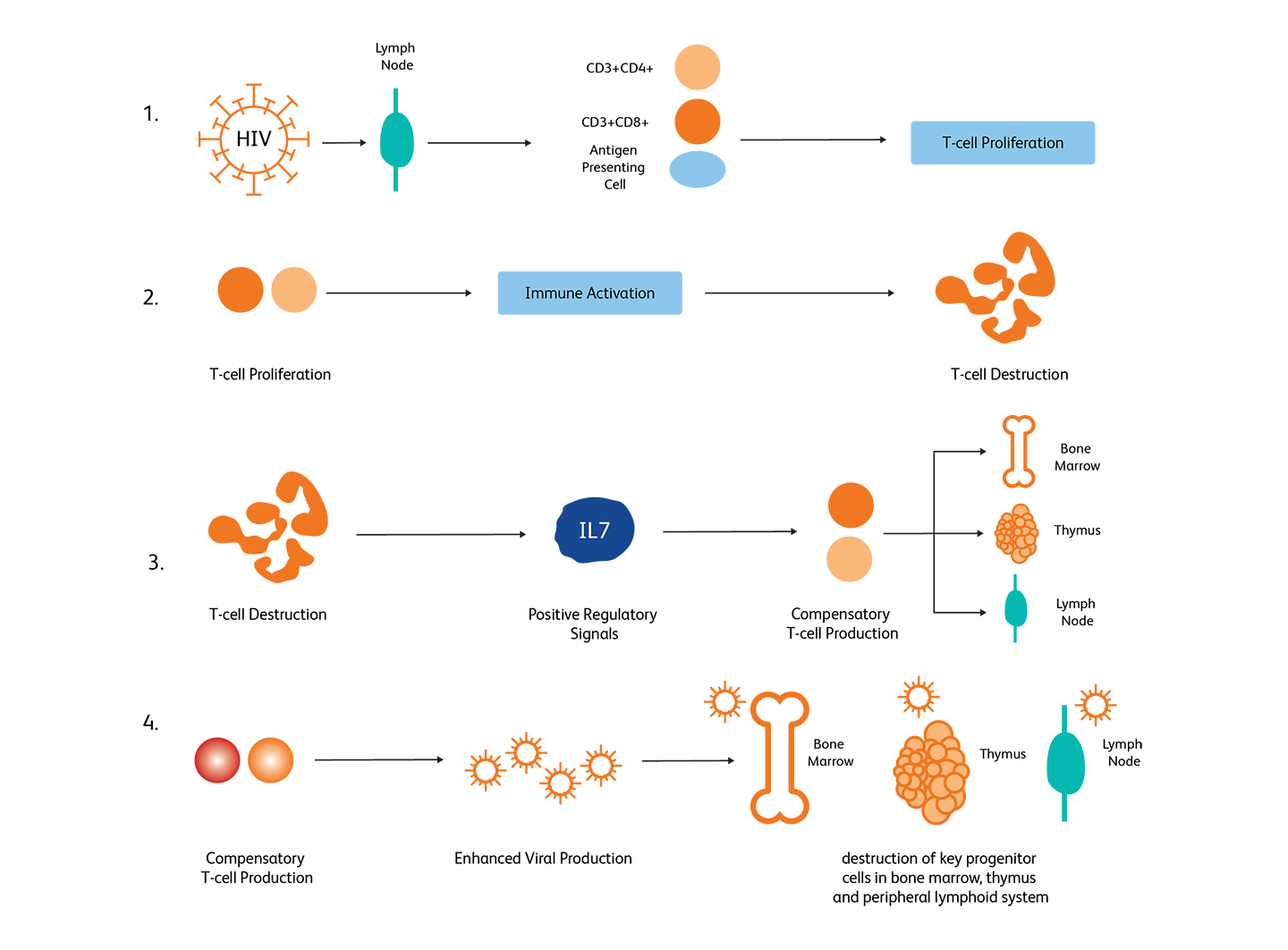
Impaired immune cell development results in most immune deficiency diseases. SCID, a PID, is a result of impaired T-cell development, leading to severe T-cell lymphopenia and lack of adaptive immune responses.2 Common variable immunodeficiency (CVID), another PID, is caused by defects in B-cell function and antibody production.3 In AIDS, a SID, HIV specifically infects and reduces the number of CD4+ subset of T lymphocytes.4

Example of immunodeficiency caused by HIV-mediated CD4+ T-cell depletion. Adopted from McCune, 2001.4
How are immune deficiencies diagnosed?
Immune disorders can be diagnosed either before or after birth depending on the condition:
Prenatal testing
These tests can be conducted using amniotic fluid, blood or cells from the chorion (future placenta).
Blood tests
Collected blood samples can be screened for counts and variety of immune cells and specifically determine the defective cells.
Genetic tests
Targeted sequencing and whole exome sequencing are also used for diagnosis of immune deficiencies, especially for diagnosing newborn SCID.5
Flow cytometry for detecting immune cell types
Flow cytometry is widely used for disease-specific assessments and immunophenotyping of immune cell populations. This evaluation help in diagnosis of immune deficiencies.
BD Biosciences clinical flow cytometry solutions, including instrumentation, software and reagents, offer the building blocks* for laboratory-developed tests used in the identification of markers associated with immune deficiencies.
In addition, the BD FACSLyric™ Flow Cytometry System when used with BD Multitest™ 6-Color TBNK Reagent + BD Trucount™ Tubes offers a clinical solution for immunological assessment of normal individuals, and patients having or suspected of having immune deficiency, with accuracy in cell phenotyping, process efficiency and standardization.
References
- Abraham RS and Aubert G. Flow cytometry, a versatile tool for diagnosis and monitoring of primary immunodeficiencies. Clin and Vaccine Immunol. 2016;23:254-271. doi: 10.1128/CVI.00001-16
- Haddad E, Logan BR, Griffith LM, et al. SCID genotype and 6-month posttransplant CD4 count predict survival and immune recovery. Blood. 2018;132(17):1737-1749. doi: 10.1182/blood-2018-03-840702
- Chinen J, Shearer WT. Advances in basic and clinical immunology 2010. J Allergy Clin Immunol. 2011;127(2):336-341. doi: 10.1016/j.jaci.2010.11.042
- McCune JM. The dynamics of CD4+ T-cell depletion in HIV disease. Nature. 2001;410:974-79. doi: 10.1038/35073648
- Richardson AM, Moyer AM, Hasadsri L, Abraham RS. Diagnostic tools for inborn errors of human immunity (primary immunodeficiencies and immune dysregulatory diseases). Curr Allergy and Asthma Rep. 2018;18(3):19. doi: 10.1007/s11882-018-0770-1
BD FACSLyric™ Flow Cytometers are Class I Laser Products.
The BD FACSLyric™ Flow Cytometer is for In Vitro Diagnostic Use with BD FACSuite™ Clinical Application for up to six colors. The BD FACSLyric™ Flow Cytometer is for Research Use Only with BD FACSuite™ Application for up to 12 colors. Not for use in diagnostic or therapeutic procedures.
*BD Biosciences clinical flow cytometry solutions, including instrumentation, software and reagents, offer the building blocks for laboratory-developed tests used in the identification of markers associated with immune deficiencies. These solutions are not FDA cleared or approved for the diagnosis of immune deficiencies. Analyte Specific Reagent. Analytical and performance characteristics are not established.
23-23019-00
Report a Site Issue
This form is intended to help us improve our website experience. For other support, please visit our Contact Us page.