
준비 및 보관
권장 분석 절차
BD® CompBeads can be used as surrogates to assess fluorescence spillover (Compensation). When fluorochrome conjugated antibodies are bound to BD® CompBeads, they have spectral properties very similar to cells. However, for some fluorochromes there can be small differences in spectral emissions compared to cells, resulting in spillover values that differ when compared to biological controls. It is strongly recommended that when using a reagent for the first time, users compare the spillover on cells and BD® CompBeads to ensure that BD® CompBeads are appropriate for your specific cellular application.
제품 고시
- Since applications vary, each investigator should titrate the reagent to obtain optimal results.
- Please refer to www.bdbiosciences.com/us/s/resources for technical protocols.
- For fluorochrome spectra and suitable instrument settings, please refer to our Multicolor Flow Cytometry web page at www.bdbiosciences.com/colors.
- Please observe the following precautions: Absorption of visible light can significantly alter the energy transfer occurring in any tandem fluorochrome conjugate; therefore, we recommend that special precautions be taken (such as wrapping vials, tubes, or racks in aluminum foil) to prevent exposure of conjugated reagents, including cells stained with those reagents, to room illumination.
- APC-Cy7 tandem fluorochrome emission is collected in a detector for fluorescence wavelengths of 750 nm and higher.
- APC-Cy7 is a tandem fluorochrome composed of Allophycocyanin (APC), which is excited by laser lines between 595 and 647 nm and serves as an energy donor, coupled to the cyanine dye Cy7™, which acts as an energy acceptor and fluoresces at 780 nm. BD Biosciences Pharmingen has maximized the fluorochrome energy transfer in APC-Cy7, thus maximizing its fluorescence emission intensity, minimizing residual emission from APC, and minimizing required electronic compensation in multilaser-laser flow cytometry systems. Note: Although every effort is made to minimize the lot-to-lot variation in residual emission from APC, it is strongly recommended that every lot be tested for differences in the amount of compensation required and that individual compensation controls are run for each APC-Cy7 conjugate.
- Warning: Some APC-Cy7 and PE-Cy7 conjugates show changes in their emission spectrum with prolonged exposure to formaldehyde. If you are unable to analyze fixed samples within four hours, we recommend that you use BD™ Stabilizing Fixative (Cat. No. 338036).
- Caution: Sodium azide yields highly toxic hydrazoic acid under acidic conditions. Dilute azide compounds in running water before discarding to avoid accumulation of potentially explosive deposits in plumbing.
- Please refer to http://regdocs.bd.com to access safety data sheets (SDS).
- Cy is a trademark of Global Life Sciences Solutions Germany GmbH or an affiliate doing business as Cytiva.
관련 제품

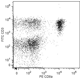
The 145-2C11 monoclonal antibody specifically binds to the 25-kDa ε chain of the T-cell receptor-associated CD3 complex that is expressed on thymocytes, mature T lymphocytes, and NK-T cells. The cytoplasmic domain of CD3e participates in the signal transduction events that activate several cellular biochemical pathways as a result of antigen recognition. Soluble 145-2C11 antibody can activate either unprimed (naive) or primed (memory/preactivated) T cells in vivo or in vitro, in the presence of Fc receptor-bearing accessory cells. In contrast, plate-bound 145-2C11 can activate T cells in the absence of accessory cells. Soluble 145-2C11 antibody has been reported to induce re-directed lysis of Fc receptor-bearing target cells by CTL clones and can also block lysis of specific target cells by antigen-specific CTL's. Under some conditions, T-cell activation by 145-2C11 antibody has been reported to result in apoptotic cell death. The 145-2C11 antibody does not cross-react with rat leukocytes. Preincubation of thymus cell suspensions at 37°C for 2-4 hours prior to staining reportedly enhances the ability of anti-CD3ε and anti-αβ TCR mAbs to detect the T-cell receptor on immature thymocytes.

개발 참고 자료 (10)
-
Beavis AJ, Pennline KJ. Allo-7: a new fluorescent tandem dye for use in flow cytometry. Cytometry. 1996; 24(4):390-395. (Methodology: Flow cytometry). 참조 보기
-
Duke RC, Cohen JJ, Boehme SA, et al. Morphological, biochemical, and flow cytometric assays of apoptosis. In: Coligan J, Kruisbeek AM, Margulies D, Shevach EM, Strober W, ed. Current Protocols in Immunology. New York: John Wiley and Sons; 1995:3.17.1-3.17.33.
-
Isakov N, Wange RL, Burgess WH, Watts JD, Aebersold R, Samelson LE. ZAP-70 binding specificity to T cell receptor tyrosine-based activation motifs: the tandem SH2 domains of ZAP-70 bind distinct tyrosine-based activation motifs with varying affinity. J Exp Med. 1995; 181(1):375-380. (Biology: Immunoprecipitation). 참조 보기
-
Kruisbeek AM, Shevach EM. Proliferative assays for T cell function. Curr Protoc Immunol. 2004; 3:3.12.1-3.12.14. (Methodology: Activation, Stimulation). 참조 보기
-
Kubo RT, Born W, Kappler JW, Marrack P, Pigeon M. Characterization of a monoclonal antibody which detects all murine alpha beta T cell receptors. J Immunol. 1989; 142(8):2736-2742. (Biology). 참조 보기
-
Leo O, Foo M, Sachs DH, Samelson LE, Bluestone JA. Identification of a monoclonal antibody specific for a murine T3 polypeptide. Proc Natl Acad Sci U S A. 1987; 84(5):1374-1378. (Immunogen: Activation, Blocking, Cytotoxicity, Immunoprecipitation, Stimulation). 참조 보기
-
Nakano H, Yamazaki T, Miyatake S, Nozaki N, Kikuchi A, Saito T. Specific interaction of topoisomerase II beta and the CD3 epsilon chain of the T cell receptor complex. J Biol Chem. 1996; 271(11):6483-6489. (Biology: Immunoprecipitation). 참조 보기
-
Portoles P, Rojo J, Golby A, et al . Monoclonal antibodies to murine CD3 epsilon define distinct epitopes, one of which may interact with CD4 during T cell activation. J Immunol. 1989; 142(12):4169-4175. (Biology: Activation, Immunoprecipitation, Stimulation). 참조 보기
-
Roederer M, Kantor AB, Parks DR, Herzenberg LA. Cy7PE and Cy7APC: bright new probes for immunofluorescence. Cytometry. 1996; 24(3):191-197. (Methodology: Flow cytometry). 참조 보기
-
Shinkai Y, Alt FW. CD3 epsilon-mediated signals rescue the development of CD4+CD8+ thymocytes in RAG-2-/- mice in the absence of TCR beta chain expression. Int Immunol. 1994; 6(7):995-1001. (Biology: Activation, Stimulation). 참조 보기
Please refer to Support Documents for Quality Certificates
Global - Refer to manufacturer's instructions for use and related User Manuals and Technical data sheets before using this products as described
Comparisons, where applicable, are made against older BD Technology, manual methods or are general performance claims. Comparisons are not made against non-BD technologies, unless otherwise noted.
For Research Use Only. Not for use in diagnostic or therapeutic procedures.
23-22944-00