-
Reagents
- Flow Cytometry Reagents
-
Western Blotting and Molecular Reagents
-
Flow Cytometry Reagents
- Immunoassay Reagents
- Single Cell Multiomics Reagents
-
Cell Preparation
-
Functional Assays
-
Microscopy and Imaging Reagents
- Western Blotting And Molecular Reagents
- Cell Preparation Separation Reagents
- Functional Cell Based Reagents
- Microscopy Imaging Reagents
- Single Cell Multiomics Reagents
- Single Cell Multinomics Reagents
-
Protocols
- BSB Protocol
-
Setting Compensation Multicolor Flow
-
Tissues Section Stain
-
Immunomicroscopy
-
Immunohistochemical
-
Immunofluorescence
-
Frozen Tissue
-
Parafin Sections
-
Fix Perm Kits
-
Protocol Direct Immunofluorscence Staining
-
Uses of Fc Block
-
Stain Lyse Wash
-
Stain Lyse No Wash
-
Mouse Splenocytes
-
Mouse Rat Leukocytes
-
Isotype Control
-
Indirect Staining Mononuclear Cells
-
Immunopurification
-
Human PBMCs
-
Human Whole Blood Samples
-
Escapee Phenomenon
-
Agarose Conjugates
-
Anti Phosphotyrosine Biotin Conjugates
-
Soluble Antibodies
-
Rabbit Polyclonal Antibodies
-
Monocloncal Antibodies
-
Horseradish Peroxidase
-
Certified Reagents
-
Biotinylated Antibodies
-
Agarose Conjugates X712261
-
Surface Staining
-
Platelet Activation
-
Intracellular Staining
-
Indirect Immunofluorescence
-
Mouse Ige
-
Cytokine Elisa
-
Induction Fas
-
Induction Dx2
-
Apoptosis By Treatment Staurosporine
-
Cell Death
-
Apo Brdu
-
Apo Direct
-
Human Cyclins
-
Detection Ki 67
-
Brdu Detection
-
Targeted mRNA Protocols
-
WTA Protocols
-
360040667732 Protocols
-
360023293831 AbSeq Protocols
-
360039007471 VDJ CDR3 Protocols
-
Annexin V Staining Protocol
-
Western Blotting with Horseradish Peroxidase Conjugates or Alkaline Phosphatase Conjugates
-
Tissue Preparation for Surface Antigen Staining
-
Account Support
-
Account FAQs
- Account FAQ Answer 1
- Account FAQ Answer 2
- Account FAQ Answer 3
- Account FAQ Answer 4
- Account FAQ Answer 5
- Account FAQ Answer 6
- Account FAQ Answer 7
- Account FAQ Answer 8
- Account FAQ Answer 9
- Account FAQ Answer 10
- Account FAQ Answer 11
- Account FAQ Answer 12
- Account FAQ Answer 13
- Account FAQ Answer 14
- Account FAQ Answer 15
- Account FAQ Answer 16
- Account FAQ Answer 21
- Create Account
- Manage Account Settings
-
PrivacyPolicy
-
Terms and Conditions
-
Account FAQs
-
- Account FAQ Answer 1
- Account FAQ Answer 2
- Account FAQ Answer 3
- Account FAQ Answer 4
- Account FAQ Answer 5
- Account FAQ Answer 6
- Account FAQ Answer 7
- Account FAQ Answer 8
- Account FAQ Answer 9
- Account FAQ Answer 10
- Account FAQ Answer 11
- Account FAQ Answer 12
- Account FAQ Answer 13
- Account FAQ Answer 14
- Account FAQ Answer 15
- Account FAQ Answer 16
- Account FAQ Answer 21
- Korea (English)
- Korea (Korea)
-
국가 / 언어 변경
Old Browser
Overview
Choose from four instrument configurations. All configurations have blue (488-nm) and violet (405-nm) lasers, which can be paired with a red (640-nm), ultraviolet (355-nm) or yellow-green (561-nm) laser, depending on your application needs.
Watch the video describing the ease of use of the BD FACSCelesta™ Flow Cytometer.

BD FACSCelesta Multicolor Flow Cytometer: Ease of Use Overview
FEATURES
The BD FACSCelesta™ Cell Analyzer enables deep and powerful insights in cell analysis at your benchtop
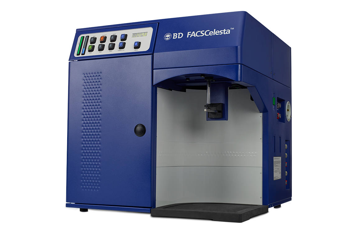
- 4 different laser configurations within a compact footprint
- Fluidics capable of acquiring tube or plate formats
- Known and trusted BD FACSDiva™ Software helps streamline flow cytometry workflows
Get more information from the BD FACSCelesta™ Cell Analyzer Brochure.
Explore the configuration sheets for different laser combinations:
BD FACSCelesta™ Flow Cytometer Configuration Sheet – BV
BD FACSCelesta™ Flow Cytometer Configuration Sheet – BVR
The BD FACSCelesta™ Cell Analyzer is optimized for use with BD Horizon Brilliant™ Dyes
More parameters mean more impact
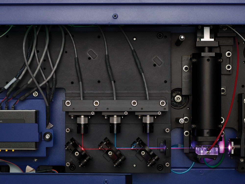
- The optical and electronics system—lasers, filters, detectors, optical paths and signal processing technologies—have been engineered for use with BD Horizon Brilliant™ Dyes.
- A system of dichroic mirrors and bandpass filters, packed into a compact polygon, splits off light from each laser and directs it to the correct detectors
- The detectors (photomultiplier tubes or PMTs) themselves fit compactly behind the filter assemblies
Options to enhance and tailor your BD FACSCelesta™ Cell Analyzer toward your specific research needs
BD FACSFlow™ Supply System (FFSS) and BD™ High Throughput Sampler (HTS) options available when you need
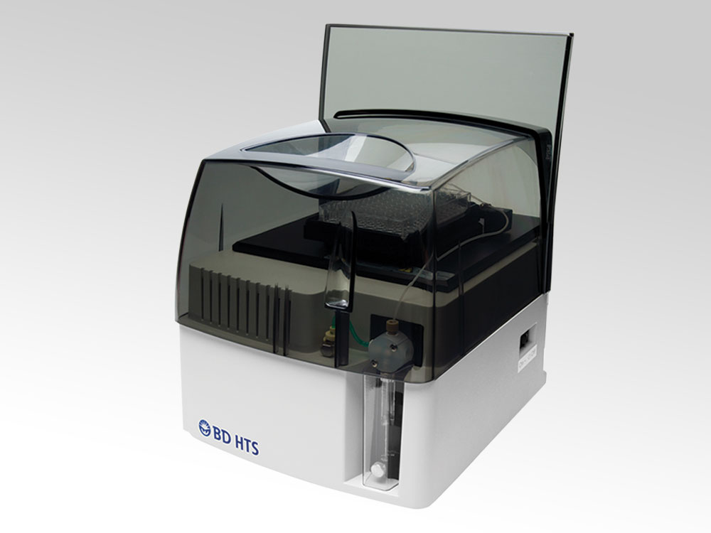
- The optional FFSS fluidics cart increases capacity and ease of use while maintaining a stable fluidics pressure and reduces daily maintenance by incorporating two 20-L containers (Cubitainers™)
- Fluidic sensors maintain constant pressure, and a fluidics monitoring system warns when sheath fluid is low or empty or when the waste container is full
- A digital control panel makes it easy to monitor the operational status of the cytometer and make adjustments
- The HTS option provides rapid, fully automated sample acquisition from microtiter plates and supports a wide range of research applications and is compatible with 96-well U, V and flat-bottom plates as well as 384-well microtiter plates
- In high-throughput mode, the HTS can process a 96-well plate in less than 15 minutes with less than 1% carryover and standard throughput mode can be selected to acquire larger sample volumes or where longer acquisition times are required
APPLICATIONS
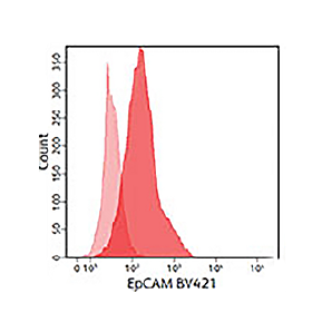
Rare cells, or cells that have few surface receptors of a marker of interest, can be difficult to detect using conventional reagents. Bright reagents are essential in resolving these dim cells from others in a sample.
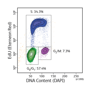
BD Horizon Brilliant™ Dyes can endow previously dim cells with much brighter fluorescence signals than traditional organic fluorescent dyes or even phycobiliproteins such as PE or APC as demonstrated in the figures below.
Optimization for these bright dyes enables the BD FACSCelesta™ System to identify cell populations with a broader range of receptor density.
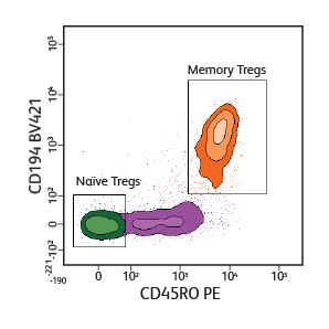
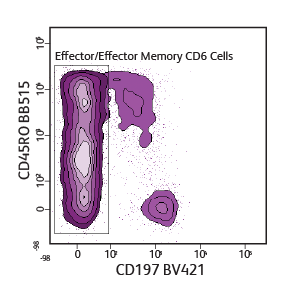
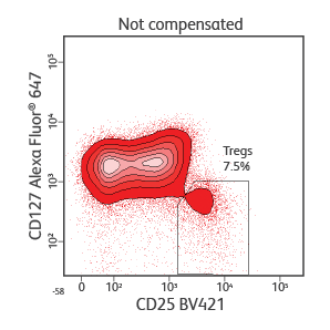
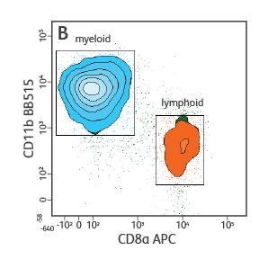
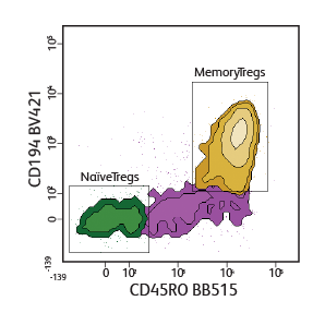
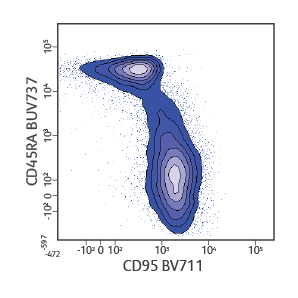
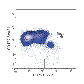
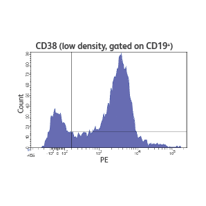
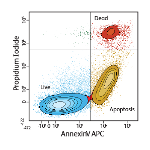
Using a broad landscape of cell function assays, flow cytometry can shed light on a variety of sample types, such as whole blood, cell lines and yeast.
Detect and analyze light scatter to measure physical characteristics of cells in suspension, such as cell shape, size and internal complexity
Add fluorescent markers to interrogate expressed or secreted proteins that reveal cell phenotype, function and status
-
Brochures
-
Filter Guide
-
Technical Specifications
-
User's Guide
-
Configuration Sheets
-
Data Book
-
Product Information Sheets
-
A flow cytometry solution for fluorescent protein analysis and beyond
-
A high-throughput flow cytometry assay to assess PD-1 receptor occupancy
-
BD FACSDiva™ Data Sheet
-
Combining instrument sensitivity with bright dyes to resolve low-density antigens
-
Designing minimal spectral overlap panels to enhance data quality
-
Enhanced cell cycle studies using multicolor flow cytometry
-
Flow cytometric assessment of CD8 + cytotoxic T-cell activation in a mixed lymphocyte reaction
-
High-dimensional flow-cytometric analysis of human B-cell populations
-
Immunophenotyping and functional analysis of rare cell populations
-
Understanding NK cell cytotoxicity against tumor cells using flow cytometry
Request a Quote
Please fill in the following information and we will get in touch with you regarding your query.
For Research Use Only. Not for use in diagnostic or therapeutic procedures.
Class 1 Laser Product.
Alexa Fluor is a trademark of Life Technologies Corporation. ATCC, HTB-22 and HTB-26 are trademarks of ATCC. CF™ is a trademark of Biotium, Inc. Cubitainer is a trademark of Hedwin Corporation. Cy is a trademark of Global Life Sciences Solutions Germany GmbH or an affiliate doing business as Cytiva. Living Colors (including mCherry dye) is a trademark of Clontech. SPHERO™ is a trademark of Spherotech, Inc.
Report a Site Issue
This form is intended to help us improve our website experience. For other support, please visit our Contact Us page.
